Section 02
Having a scan
The referral, the checks a department carries out, what contrast agents are, and the route a report takes back to the clinician who asked for it.
Every examination that uses ionising radiation in the United Kingdom follows the same broad pattern. A written request arrives from an authorised health professional, the imaging department carries out a set of checks required by law, the scan takes place, and a recorded evaluation travels back to whoever made the request. This page outlines that shared journey and points to the two pages that follow it in this section.
Four stages, one framework
The first stage is the referral. Under IR(ME)R 2017, a referrer is defined as a person "entitled in accordance with the employer's procedures to refer individuals for exposure to a practitioner", and who is "a registered health care professional", according to the definition given in the regulations on legislation.gov.uk. The referrer must supply sufficient medical data, such as previous diagnostic information or medical records, so that a practitioner can decide whether the exposure offers a sufficient net benefit. A request is therefore never an automatic order: the practitioner is responsible for the justification of each exposure.
The second stage is a set of checks performed by the service itself. The employer must maintain a written procedure, described in Schedule 2 of IR(ME)R 2017, for making enquiries of individuals of childbearing potential to establish whether the individual is or may be pregnant or breastfeeding. The same Schedule requires a procedure ensuring that, wherever practicable and prior to an exposure, the individual to be exposed or their representative is provided with adequate information relating to the benefits and risks associated with the radiation dose. For magnetic resonance imaging, the NHS page dated 11 September 2025 describes hospitals using a questionnaire covering metal implants, citing surgical clips, a pacemaker, dental fillings or bridges, and asking patients not to wear anything containing metal, such as jewellery, a bra, hair clips or hearing aids.
The examination itself
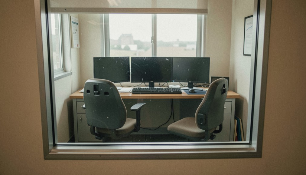
During the scan, the practical aspects are the responsibility of the operator, who must comply with the employer's procedures under IR(ME)R 2017. The radiographer often works from a neighbouring room: the NHS MRI page dated 11 September 2025 describes the radiographer operating the scanner from another room while keeping contact with the patient. Some examinations involve a substance given to improve the images, and the page on contrast agents explains what may be injected or swallowed. The NHS CT page dated 08 November 2023 notes that contrast can be given by drink, injection or rectally, and mentions passing sensations such as feeling hot and flushed or a metal taste in the mouth. The legal side of a request is covered in the referral.
The recorded evaluation
The final stage is the report. Schedule 2 of IR(ME)R 2017 requires the employer to have a procedure for the carrying out and recording of an evaluation for each exposure, including, where appropriate, factors relevant to patient dose. The Royal College of Radiologists describes radiologists as specialist doctors trained to read and interpret images across X-ray, ultrasound, CT, MRI and PET. For ultrasound, the NHS page dated 25 February 2025 states that the doctor who requested the scan receives the conclusions, and that a follow-up appointment may be needed to discuss them. The NHS reports result timescales too: MRI results usually take between 1 and 2 weeks, and CT results usually take between 1 and 2 weeks but can take up to 4 weeks.
The other doors of the publication: Scans explained, Radiation and safety and People and history. Every one of them points back to modalities at a glance.
In this section
10 pages, newest first.
-
The referral
No imaging examination starts with the person having it. It starts with a written request from a clinician, and in the UK that request has a legal weight.
6 September 2026
-
Contrast agents
A contrast agent does not make a picture. It changes how a tissue or a vessel appears in a picture that would have been taken anyway.
6 September 2026
-
The route of a request
A scan is the middle of a journey, not the start of one. The request, the justification, the exposure and the report each have their own rule and their own owner.
6 September 2026
-

The American bill
The same examination, a different paper trail. In the United States a scan can arrive afterwards as two separate bills, and a body of federal rules exists to govern what may be charged.
6 September 2026
-
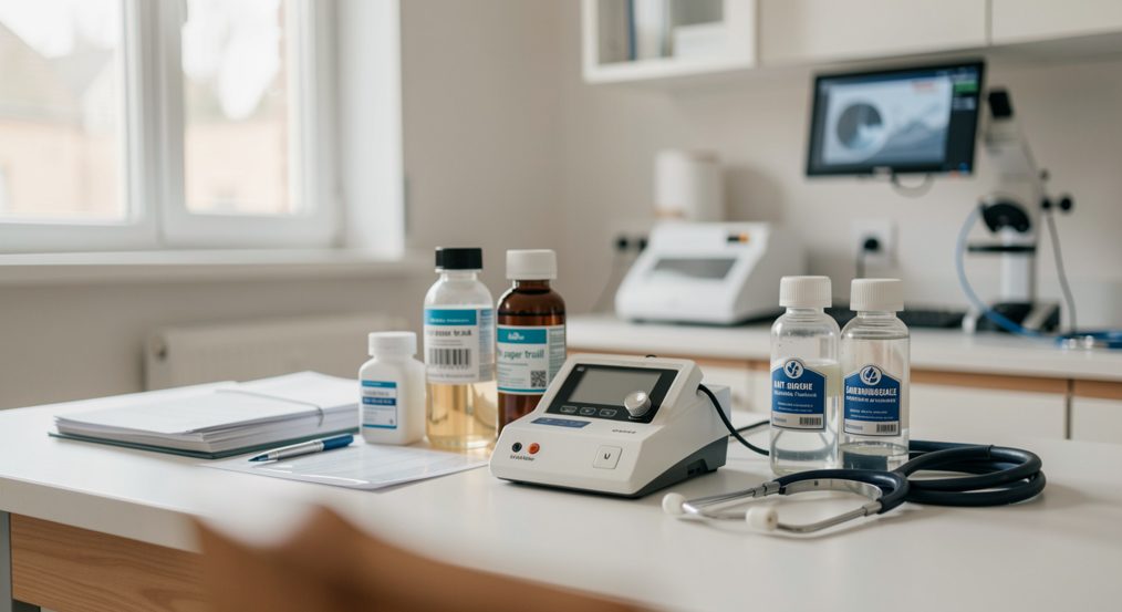
The paper trail
A scan produces an archive as well as an image. Most of that trail is compulsory under the 2017 regulations, and each document in it answers to a written duty.
6 September 2026
-
Where the pictures go
The pictures stay; the report travels. The images go into the department’s archive under record-keeping duties, while the interpretation makes the journey back to whoever asked the question.
6 September 2026
-

Breath Meditation: A Beginner's First Sitting
A plain guide to starting breath meditation: posture, counting the breath, how long a first sitting lasts, and keeping a daily practice going between retreats.
6 September 2026
-

A First Visit to a Family Practice: Records and Routines
What to bring to a first visit at a family practice, how an annual physical is organised, and when a patient portal replaces a telephone call.
6 September 2026
-
Entering a Care Home in France: What Families Face
How a place in a French EHPAD is chosen, what the first days involve, the contract of residence, the GIR grid and the financial help families can claim.
6 September 2026
-
The First Year of Fatherhood: Leave and Paperwork
How paternity and parental leave work in France, the paperwork with the employer and the health insurance fund, and the everyday questions of a baby's first
6 September 2026