Medical imaging, explained without advice
Inside the Scanner
An imaging examination is a picture taken to answer a question a clinician has already asked. This site explains what each kind of picture is, how it is made, and who decides that it should be made at all.
- 2.7 mSv a year, UK average Natural background dose
- 0.014 mSv for a chest X-ray From the same UKHSA table
- 4 Duty holders in law Referrer, practitioner, operator, employer
- 1897 The Roentgen Society The first X-ray association
The two dose figures are read in the UK Health Security Agency comparison table Ionising radiation: dose comparisons, published 18 March 2011. The four duty holders are defined in regulation 2 of the Ionising Radiation (Medical Exposure) Regulations 2017 on legislation.gov.uk. The 1897 date is published by the Royal College of Radiologists. All four were read on 6 September 2026. The same figures are published in modalities at a glance.
The four sections
From the methods themselves to the rules that govern their use.
-
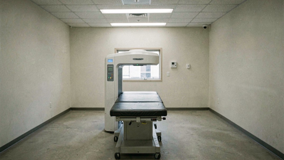
Scans Explained: X-ray, CT, MRI, Ultrasound
Five methods, five different physical measurements, and the reason no single scan answers every question.
Section 01
-
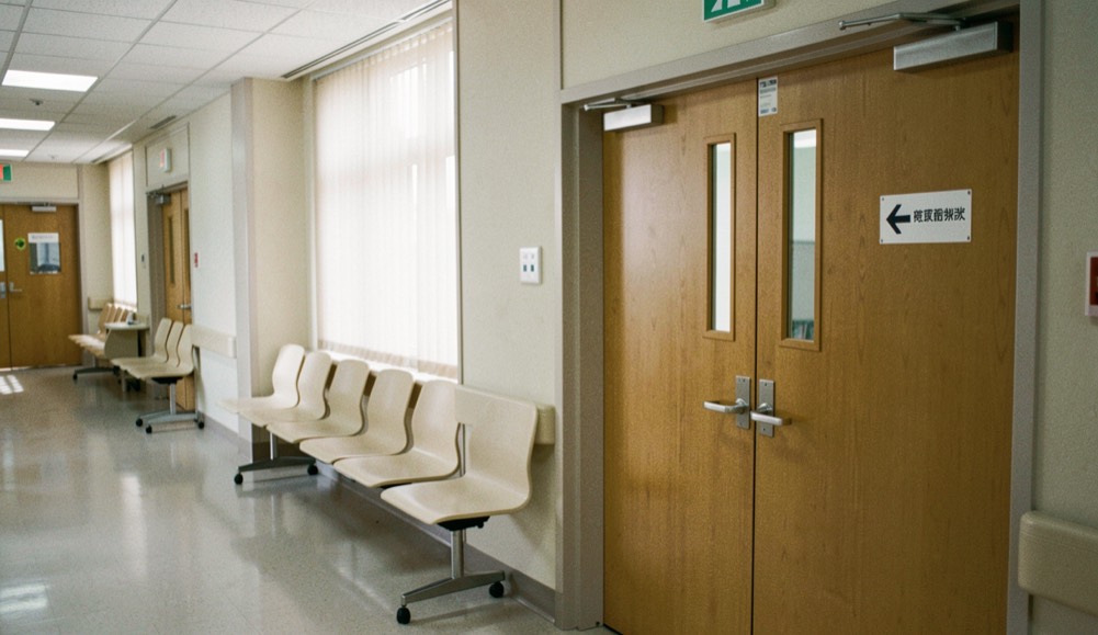
Having a Scan: How the Appointment Runs
A request, a set of checks, an examination and a report: the four steps that every imaging appointment shares.
Section 02
-
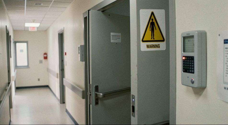
Radiation and Safety in Medical Imaging
Every exposure has to be justified before it is optimised, and both words have a legal meaning in the UK.
Section 03
-
Who Does What in an Imaging Department
Different jobs, different registers, and a discipline barely older than photography of the living body.
Section 04
The two standing references
Cited by every section, each row carrying the source it was read from.
-
Modalities at a glance
The comparison table: what each method measures, and where that was read.
Reference
-
Every page on this site
The full list, with the date of the work that produced each one.
-
Having an MRI scan
Magnetic resonance
Magnetic resonance imaging makes pictures from radio signals given off by the body inside a strong magnetic field. It uses no ionising radiation, and that single fact shapes everything else about it.
-
CT scans
Computed tomography
A CT scanner is an X-ray tube that travels around the body while a ring of detectors records what gets through. The picture is calculated, not photographed.
-
Ultrasound
Sonography
Ultrasound sends pulses of high frequency sound into the body and listens for the echoes. Nothing is emitted that the body has not already been exposed to in kind.
-
Plain X-rays
Radiography
A radiograph is a shadow. What reaches the detector is what the beam was not stopped by, which is why bone is white and air is black.
-
The referral
The request
No imaging examination starts with the person having it. It starts with a written request from a clinician, and in the UK that request has a legal weight.
-
Contrast agents
Contrast media
A contrast agent does not make a picture. It changes how a tissue or a vessel appears in a picture that would have been taken anyway.
-
Radiation doses
Dose
Dose figures are only meaningful next to the source they were published in and the year that source was written. Every number on this page carries both.
-
IR(ME)R 2017
Regulation
Referrer, practitioner, operator, employer. Four defined roles decide whether a medical exposure happens at all, and each one carries a duty that is written down.
-
Who does what
The professions
The person who positions you is very unlikely to be the person who writes the report. In the UK the two jobs sit on different registers and follow different training routes.
-
The British institutions
Institutional history
The history of the machines is told everywhere and sourced almost nowhere. The history of the British bodies that govern their use is published by those bodies themselves, with dates.
-
Magnetic fields, not X-rays
Magnetic resonance
An MRI scanner makes its pictures from magnetic fields and the signals the body returns inside them. No ionising radiation is involved, and that fact organises everything else about the examination.
-
Attenuation
Computed tomography
Attenuation is the word for what tissue does to the beam: how much of it is stopped on the way through. A CT report turns that stopping into a number.
-
When the patient is an animal
Beyond the patient
The physics of a scan does not change with the species on the table. The paperwork does, because the regulations this site covers are written for human patients.
-
The route of a request
The journey
A scan is the middle of a journey, not the start of one. The request, the justification, the exposure and the report each have their own rule and their own owner.
-
The American bill
Another system
The same examination, a different paper trail. In the United States a scan can arrive afterwards as two separate bills, and a body of federal rules exists to govern what may be charged.
-
The paper trail
The record
A scan produces an archive as well as an image. Most of that trail is compulsory under the 2017 regulations, and each document in it answers to a written duty.
-
Where the pictures go
The archive
The pictures stay; the report travels. The images go into the department’s archive under record-keeping duties, while the interpretation makes the journey back to whoever asked the question.
-
What the scan room is made of
The room
An X-ray room is built, not just furnished. The door, the sign and the screen are each the physical form of a written duty.
-
Does a dose add up?
Dose
Every exposure has a figure, and the figures can be summed. What the sum means is the harder question, and the regulations answer it exposure by exposure.
-
Reading an image
The craft
Reading is the trained act that turns a picture into a report. In the UK the people who acquire the image and the people who read it sit on different registers.
-
How imaging got its names
The vocabulary
Every method is named for the thing it measures. The professional titles, the dose quantities and the legal roles each have a register of names of their own.
-
The software in the room
The systems
A modern scanner is a computer wrapped in a machine. The image is calculated, the record is stored, and the regulations assume the software works.
-
How pages are checked
The method
Every page of substance is re-read against the sources it names before it is published. This page describes the check, and the same discipline applied elsewhere.
-
Infection Control in Shops: A Written Policy
Radiation and safety
How small businesses write a basic infection control policy, manage staff sickness and return to work, and word the notices customers actually read.
-
Breath Meditation: A Beginner's First Sitting
Having a scan
A plain guide to starting breath meditation: posture, counting the breath, how long a first sitting lasts, and keeping a daily practice going between retreats.
-
A First Visit to a Family Practice: Records and Routines
Having a scan
What to bring to a first visit at a family practice, how an annual physical is organised, and when a patient portal replaces a telephone call.
-
Dutch Basic Insurance and Physiotherapy
People and history
How the Dutch basic health insurance covers physiotherapy, what the Besluit zorgverzekering defines, and why the decree prints no amounts.
-
Body Composition and Daily Habits
Scans explained
A plain English look at what the body is made of and how water, magnesium, fibre, sleep, cortisol and movement after forty change it over time.
-
Family Constellations: Method and Evidence
People and history
What happens at a family constellation seminar, what the method claims, what evidence and professional criticism say, and where to turn if it unsettles.
-
Entering a Care Home in France: What Families Face
Having a scan
How a place in a French EHPAD is chosen, what the first days involve, the contract of residence, the GIR grid and the financial help families can claim.
-
The First Year of Fatherhood: Leave and Paperwork
Having a scan
How paternity and parental leave work in France, the paperwork with the employer and the health insurance fund, and the everyday questions of a baby's first
-
Legal Hemp and CBD in France: What the Rules Say
Radiation and safety
A plain guide to legal hemp and CBD in France: the THC threshold, European case law, food and animal status, and what the documented uses show.
-
Diclofenac and Anti-Inflammatory Medicines
Radiation and safety
What diclofenac does, how the 50 mg dose and gel differ from tablets, how side effects are reported, and how paracetamol differs from an anti-inflammatory.
Six pages to begin with
One scan, one appointment, one set of figures and one set of rules.
-
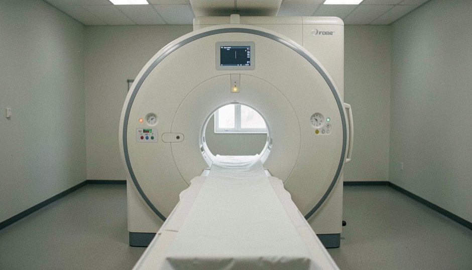
Having an MRI scan
Magnetic resonance imaging makes pictures from radio signals given off by the body inside a strong magnetic field. It uses no ionising radiation, and that single fact shapes everything else about it.
6 September 2026
-
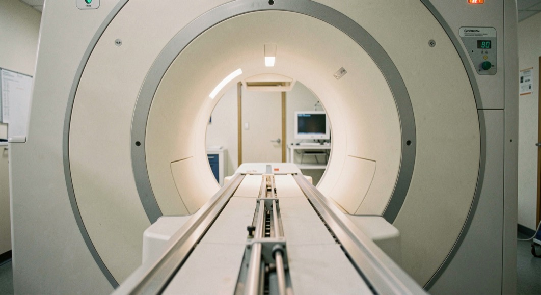
CT scans
A CT scanner is an X-ray tube that travels around the body while a ring of detectors records what gets through. The picture is calculated, not photographed.
6 September 2026
-
The referral
No imaging examination starts with the person having it. It starts with a written request from a clinician, and in the UK that request has a legal weight.
6 September 2026
-
Radiation doses
Dose figures are only meaningful next to the source they were published in and the year that source was written. Every number on this page carries both.
6 September 2026
-
IR(ME)R 2017
Referrer, practitioner, operator, employer. Four defined roles decide whether a medical exposure happens at all, and each one carries a duty that is written down.
6 September 2026
-
Who does what
The person who positions you is very unlikely to be the person who writes the report. In the UK the two jobs sit on different registers and follow different training routes.
6 September 2026
Inside the Scanner explains what happens during an imaging examination: how it is requested, how it is carried out and how it is reported. Every figure is taken from a named primary source and dated, and the checking method is set out in sources and method.
Five more reading rooms
Independent publications that follow the same rule as this one: every claim next to the source it was read from.
-

Low Altitude Review
A reading site on aerial photography made between roughly thirty and five hundred feet, with the lift arithmetic and the US rules for moored balloons printed beside it.
Reading room
-
the unpaid invoice guide
An editorial guide to business-to-business unpaid invoices: evidence files, reminder sequences, and the escalation rules across the UK, the US and the EU.
Reading room
-
pipeline incident records
A documentary site on US pipeline safety data: incident records by system, the causes named in official filings, and the 49 CFR framework each figure answers to.
Reading room
-
independent film production
A blog about making short and medium-length independent films: shoot planning, small crews, portable light, and the route into festivals.
Reading room
-
Tadcaster war memorials
A local history site on the war memorials of Tadcaster and the First World War records behind the names, from dedication ceremonies to pension files.
Reading room
-
The Prevention Desk
The Prevention Desk is an independent English-language magazine covering infection prevention in shops, workplaces and public spaces, with practical notes on ventilation, cleaning and a small business infection control plan for everyday premises.
Reading room
-
MDR1 Gene Guide
In a different field, the MDR1 gene guide sets out the MDR1 mutation in dogs, the drugs that can harm herding breeds, and the screening, veterinary and daily-care questions owners and breeders bring to it.
Reading room
-
Zweetprobleempjes
A Dutch information site, everyday sweating and hyperhidrosis, sets out causes and daily measures by body area, clothing and visible protection, and the difference between deodorant and antiperspirant, for adults troubled by sweat at work or in company.
Reading room