Section 01
Scans explained
What each imaging method actually measures, what kind of picture it produces, and which questions it is used to answer in UK practice.
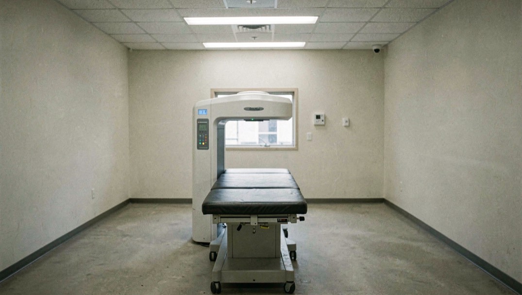
Medical imaging is described by the UK Health Security Agency in its guide published on 22 September 2022 as "one of the biggest advances in modern medicine", used to help diagnose and treat patients. The guide covers five broad families of examination: X-ray, CT, ultrasound, MRI and nuclear medicine imaging. Behind that list sits a fundamental divide, and understanding it is the clearest way into the whole subject.
Two families, divided by physics
According to the UK Health Security Agency, the X-ray family and nuclear medicine both use ionising radiation. The X-ray family is wide: general radiography, which produces 2D images; dental radiography, described by the same source as the most common type of X-ray examination; fluoroscopy, which produces moving images of parts of the body; interventional radiology, which uses X-rays to produce moving images that can be used to guide and deliver treatment; and CT, which produces 3D images of the body. Nuclear medicine, including PET-CT and PET-MRI, uses a small amount of a radioactive substance that is usually injected into a vein, as the UK Health Security Agency explains.
On the other side of the divide, ultrasound and MRI are described by the UK Health Security Agency as "types of imaging that do not use X-rays or radioactive substances". Ultrasound works with sound waves, and MRI is carried out in "a large machine with a short tunnel through it, and a flat bed that moves through the scanner" (NHS, 11 September 2025).
Different machines, different questions
The NHS describes an X-ray as a test that takes pictures of the inside of the body using a small amount of radiation, with bones, teeth, lungs and the digestive system among the targets it names. A CT scan, the NHS says, takes detailed pictures of the inside of the body, using a large ring that moves around part of the body as the person lies on a flat bed. An MRI scanner is described by the NHS as a large machine with a short tunnel through it and a flat bed that moves through the scanner, and the examination usually takes between 15 minutes and 1 hour. Ultrasound, again according to the NHS, is used to see images of the inside of the body, such as muscles, organs, or a baby in the womb, and the scan lasts around 10 to 45 minutes.
Because the underlying measurement differs, the choice between methods depends on what is being looked at, and that comparison is easier to see on the modalities table, which sets the main techniques side by side on a single grid.
What this section covers
This section of Inside the Scanner contains four pages, one for each of the principal methods:
- Plain X-rays, covering general and dental radiography, fluoroscopy and interventional radiology.
- CT scans, the technique that uses X-rays to build 3D images of the body.
- Ultrasound, the method based on sound waves rather than radiation.
- Having an MRI scan, the method that relies on magnetic resonance rather than X-rays or radioactive substances.
Each page describes how the machine works, what the NHS says the examination involves, and how long it typically lasts, so that the physics and the practical experience of the scan can be read together.
The other doors of the publication: Having a scan, Radiation and safety and People and history. Every one of them points back to modalities at a glance.
In this section
8 pages, newest first.
-
Having an MRI scan
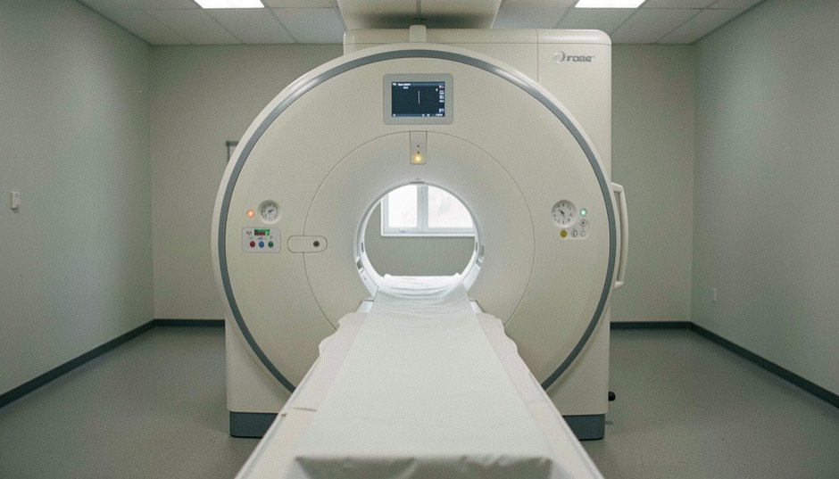
Magnetic resonance imaging makes pictures from radio signals given off by the body inside a strong magnetic field. It uses no ionising radiation, and that single fact shapes everything else about it.
6 September 2026
-
CT scans
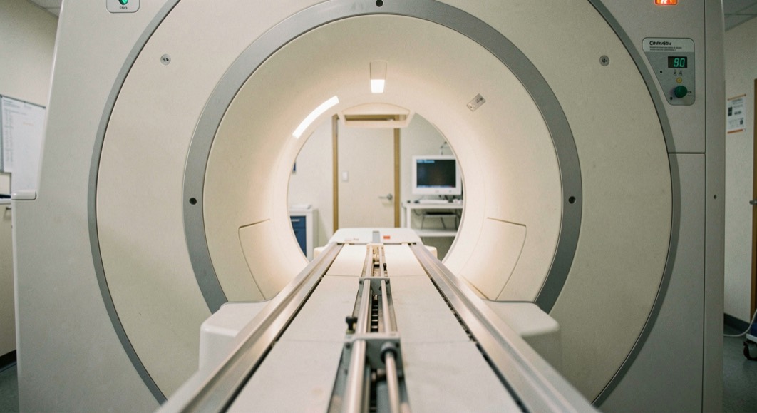
A CT scanner is an X-ray tube that travels around the body while a ring of detectors records what gets through. The picture is calculated, not photographed.
6 September 2026
-
Ultrasound
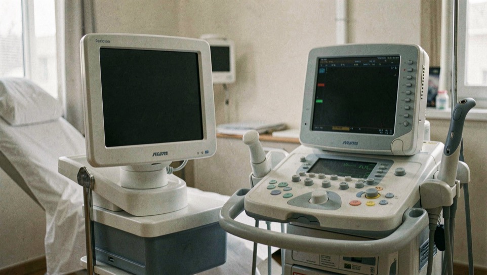
Ultrasound sends pulses of high frequency sound into the body and listens for the echoes. Nothing is emitted that the body has not already been exposed to in kind.
6 September 2026
-
Plain X-rays
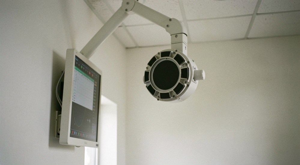
A radiograph is a shadow. What reaches the detector is what the beam was not stopped by, which is why bone is white and air is black.
6 September 2026
-
Magnetic fields, not X-rays
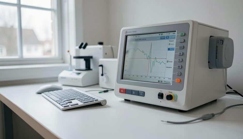
An MRI scanner makes its pictures from magnetic fields and the signals the body returns inside them. No ionising radiation is involved, and that fact organises everything else about the examination.
6 September 2026
-

Attenuation
Attenuation is the word for what tissue does to the beam: how much of it is stopped on the way through. A CT report turns that stopping into a number.
6 September 2026
-

When the patient is an animal
The physics of a scan does not change with the species on the table. The paperwork does, because the regulations this site covers are written for human patients.
6 September 2026
-

Body Composition and Daily Habits
A plain English look at what the body is made of and how water, magnesium, fibre, sleep, cortisol and movement after forty change it over time.
6 September 2026